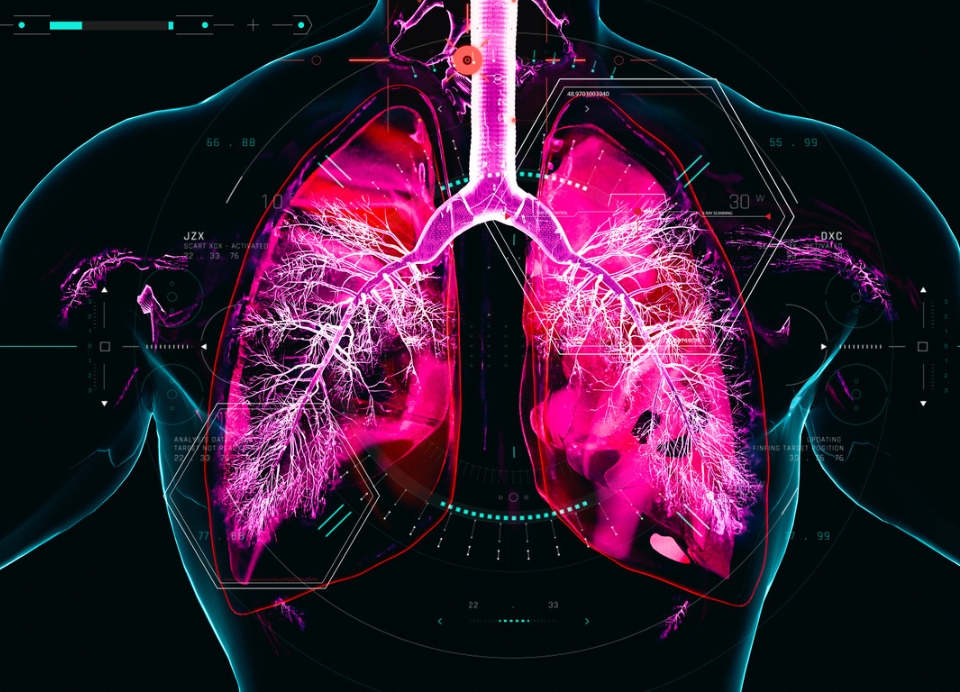
The COVID-19 pandemic demonstrated the urgent need for better understanding of how viruses interact with our lungs.
That’s the focus of the Doherty Institute’s Associate Professor Ash Haque and his team’s work: using a new technology called spatial transcriptomics – which enables researchers to look at almost every single cell type within an organ – they’ll create a detailed model of human lungs at the cellular level.
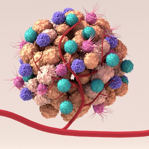
Spatial transcriptomics is a revolutionary technology that allows scientists to examine and interpret thousands of possible cell interactions and understand nuanced characteristics within the tissue they naturally reside in. It enables understanding of cell types and cellular interaction at an entirely new level: the data sets available now are exponentially more advanced than what we had just a few years ago.
“Thanks to technology advancement over recent years, we’ve seen a quantum leap in our understanding of cellular interaction. Where we could previously 'ask' a cell what it might do in 10 different situations, we can now ask questions for thousands of possible scenarios,” explains Associate Professor Haque.
“This level of detail can give you clues as to why cells behave in certain ways and point to new applications for existing medicines or new therapies. Cells behave differently depending on the tissue they're in, so understanding more about cells in their natural context rather than a Petri dish gives us critical insights into how the lungs respond during infection.”
The lungs are particularly complex structures and comprising different cell types with distinct functions. Understanding how this cellular community behaves during viral infection is critical to developing targeted therapeutics for viruses of pandemic potential.
“Our vision is to create detailed visual maps that transform our understanding of lung immunity during infection, which will help identify new therapeutic targets to prevent severe disease during respiratory pandemics,” says Associate Professor Haque.
By providing clearer predictions of cellular responses to infection, this research could also significantly improve the success rate of clinical trials for new therapeutics.
The team also wants to help give people a greater understanding of lungs and how they work, so it’s working with design and augmented reality experts to create educational 3D tools to show students the lung models.
“Once you start to see all these cells interacting, it helps people understand how the body functions. So we're creating visual tools that show people the beauty and magnificence of their immune system in action.
“I’m particularly excited by this project because it combines different expertise to progress our understanding of immunity. Traditional medical laboratory scientists joining forces with computational experts who can make sense of the enormous datasets generated, all for the greater good of humanity,” Associate Professor Haque concluded.
Project title: Mapping the cellular interactome of the lung
Chief Investigator: Ashraful Haque
Co-Investigators: Associate Professor Linda Wakim, Professor Scott Mueller, Dr Amanda Oliver and Dr Jan Schroeder.
Spotlight on Therapeutics: This content series profiles the projects and people behind the Cumming Global Centre for Pandemic Therapeutics innovative research.
More updates and news from the Doherty Institute